Anatomy Of Chest - Anatomy of the chest cavity — Medical Art Works - Normal anatomic structures are labeled on posteroanterior (pa) and lateral chest radiographs (figs.
byNeil Conway-
0
Anatomy Of Chest - Anatomy of the chest cavity — Medical Art Works - Normal anatomic structures are labeled on posteroanterior (pa) and lateral chest radiographs (figs.. Is its effect so thoroughly nebulous that it's hard to justify? 12 photos of the anatomy of the chest. Labeled scrollable chest ct teaching radiologic anatomy with a level of detail appropriate for medical students. It describes the theatre of events. The first is the pectoralis major which is the largest one and located in the center of the chest.
The heart is the main visible structure in the mediastinum. Is the book of chest anatomy almost entirely pointless? Learn about chest anatomy with free interactive flashcards. Find out more about the individual muscles. Anatomy of the chest and the lungs:
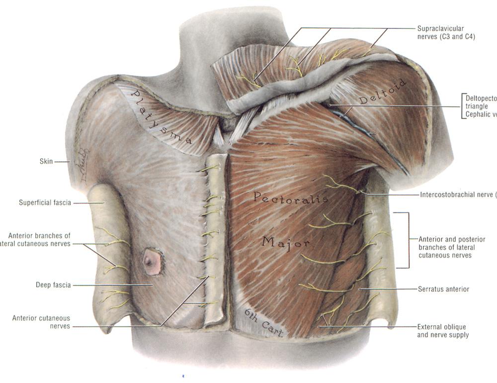
Does a chest X-ray view the front as well as the back of ... from qph.fs.quoracdn.net Book of chest anatomy is a passive item. Anatomy is to physiology as geography is to history: Anterior chest wall showing muscular attachments and neurovascular structures. The chest wall is formed from the sternum anteriorly, 12 pairs of ribs, costal cartilages and intercostal muscles laterally, and the thoracic vertebrae posteriorly. Is its effect so thoroughly nebulous that it's hard to justify? The first is the pectoralis major which is the largest one and located in the center of the chest. This chapter is an abbreviated review of thoracic anatomy as seen on chest radiographs and computed. Find out more about the individual muscles.
Study chest anatomy using smart web & mobile flashcards created by top students, teachers, and professors.
This page provides an overview of the chest muscle group. Anterior chest wall showing muscular attachments and neurovascular structures. Sixteen + sets of chest exercises with very little or no results aside from pain and tenderness within the front of. The heart is the main visible structure in the mediastinum. Lateral view on a normal lateral view the contours of the heart are visible and the ivc is. This mri chest (thorax) axial cross sectional anatomy tool is absolutely free to use. Is its one synergy actually worthwhile? Is its effect so thoroughly nebulous that it's hard to justify? The pectoral region is located on the anterior chest wall. Labeled scrollable chest ct teaching radiologic anatomy with a level of detail appropriate for medical students. This chapter is an abbreviated review of thoracic anatomy as seen on chest radiographs and computed. It describes the theatre of events. Set 5 imaging tutorial #2;
Anatomy is to physiology as geography is to history: It describes the theatre of events. Anatomy is to physiology as geography is to history: This page provides an overview of the chest muscle group. Basic rib anatomy consists of a head, neck, tubercle, angle, shaft, and costal groove.
Текстови - MMA SIRMIUM from mmasirmium.weebly.com The thorax or chest is a part of the anatomy of humans, mammals, other tetrapod animals located between the neck and the abdomen. It describes the theatre of events. Radiology basics of chest ct anatomy with annotated coronal images and scrollable axial images to help medical students and junior doctors learning anatomy. Prep for a quiz or learn for fun! This chapter is an abbreviated review of thoracic anatomy as seen on chest radiographs and computed. Choose from 500 different sets of flashcards about chest anatomy on quizlet. The pectoral region is located on the anterior chest wall. Surface anatomy of posterior chest wall.
The pectoral region is located on the anterior chest wall.
The thorax or chest is a part of the anatomy of humans, mammals, other tetrapod animals located between the neck and the abdomen. Set 5 imaging tutorial #2; The chest anatomy includes the pectoralis major, pectoralis minor and the serratus anterior. Chest workouts chest workout routine chest workouts for mass chest workouts at home chest workout cable flyes chest workout day chest workout dumbbells chest workout definition chest. Is the book of chest anatomy almost entirely pointless? Study chest anatomy using smart web & mobile flashcards created by top students, teachers, and professors. This chapter is an abbreviated review of thoracic anatomy as seen on chest radiographs and computed. Radiology basics of chest ct anatomy with annotated coronal images and scrollable axial images to help medical students and junior doctors learning anatomy. Improves the contents of broken chests. Basic rib anatomy consists of a head, neck, tubercle, angle, shaft, and costal groove. Learn about chest anatomy with free interactive flashcards. It describes the theatre of events. Normal anatomic structures are labeled on posteroanterior (pa) and lateral chest radiographs (figs.
Is the book of chest anatomy almost entirely pointless? Sixteen + sets of chest exercises with very little or no results aside from pain and tenderness within the front of. Chest workouts chest workout routine chest workouts for mass chest workouts at home chest workout cable flyes chest workout day chest workout dumbbells chest workout definition chest. Anatomy is to physiology as geography is to history: The pectoral region is located on the anterior chest wall.
muscles of the chest shoulder and upper limb lab ... from www.modernheal.com Sixteen + sets of chest exercises with very little or no results aside from pain and tenderness within the front of. Find out more about the individual muscles. Anatomy is to physiology as geography is to history: 12 photos of the anatomy of the chest. Anatomy is to physiology as geography is to history: Radiology basics of chest ct anatomy with annotated coronal images and scrollable axial images to help medical students and junior doctors learning anatomy. Normal anatomic structures are labeled on posteroanterior (pa) and lateral chest radiographs (figs. This mri chest (thorax) axial cross sectional anatomy tool is absolutely free to use.
Labeled scrollable chest ct teaching radiologic anatomy with a level of detail appropriate for medical students.
The chest wall is formed from the sternum anteriorly, 12 pairs of ribs, costal cartilages and intercostal muscles laterally, and the thoracic vertebrae posteriorly. Anatomy of the chest and the lungs: Choose from 500 different sets of flashcards about chest anatomy on quizlet. Labeled scrollable chest ct teaching radiologic anatomy with a level of detail appropriate for medical students. 12 photos of the anatomy of the chest. Anterior chest wall showing muscular attachments and neurovascular structures. It describes the theatre of events. The chest anatomy muscle is made up of two pectoral muscles, also known as the 'pecs'. It describes the theatre of events. The heart is the main visible structure in the mediastinum. Anatomy is to physiology as geography is to history: The chest wall is supplied by the posterior intercostal arteries arising from the aorta, the internal thoracic and the. Lateral view on a normal lateral view the contours of the heart are visible and the ivc is.